Prof. CaoXia Shenyang Campo Imaging Diagnostic Center
Precision 32 spectrum CT is the first CT product independently developed by Campo Imaging Medical. Unique 0.275mm ultra-thin precision CT image, sub-millimeter scanning in a large coverage area, which not only shorts the scanning time, but also has higher accuracy, and the subject receives lower radiation dose. It can clearly display the peripheral bronchi, pulmonary vessels, small lesions in the lung and related adjacent relationships, etc. It has unique value in the diagnosis of bronchiectasis, emphysema, lung infection and small pulmonary nodules.
Inspection method: Conventional scanning, 1.1mm reconstruction, 0.275mm precision scanning and MPR reconstruction were performed for targeted lesions.
Case 1
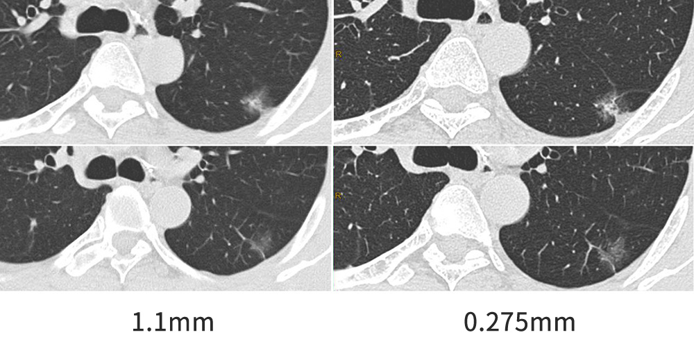
Male, 57 years old, Infiltrating adenocarcinoma of the inferior lobe of left lung (IAC)
1.1mm reconstruction showed ground glass density under the pleura of left lower lobe dorsal segment. The 0.275mm thin scanning showed solid components in the center and ground glass density shadow around it. The relationship between the shadow of adjacent blood vessels and adjacent pleural traction were more clear, and the benign and malignant diagnosis of lesions was more clear.

Case 2
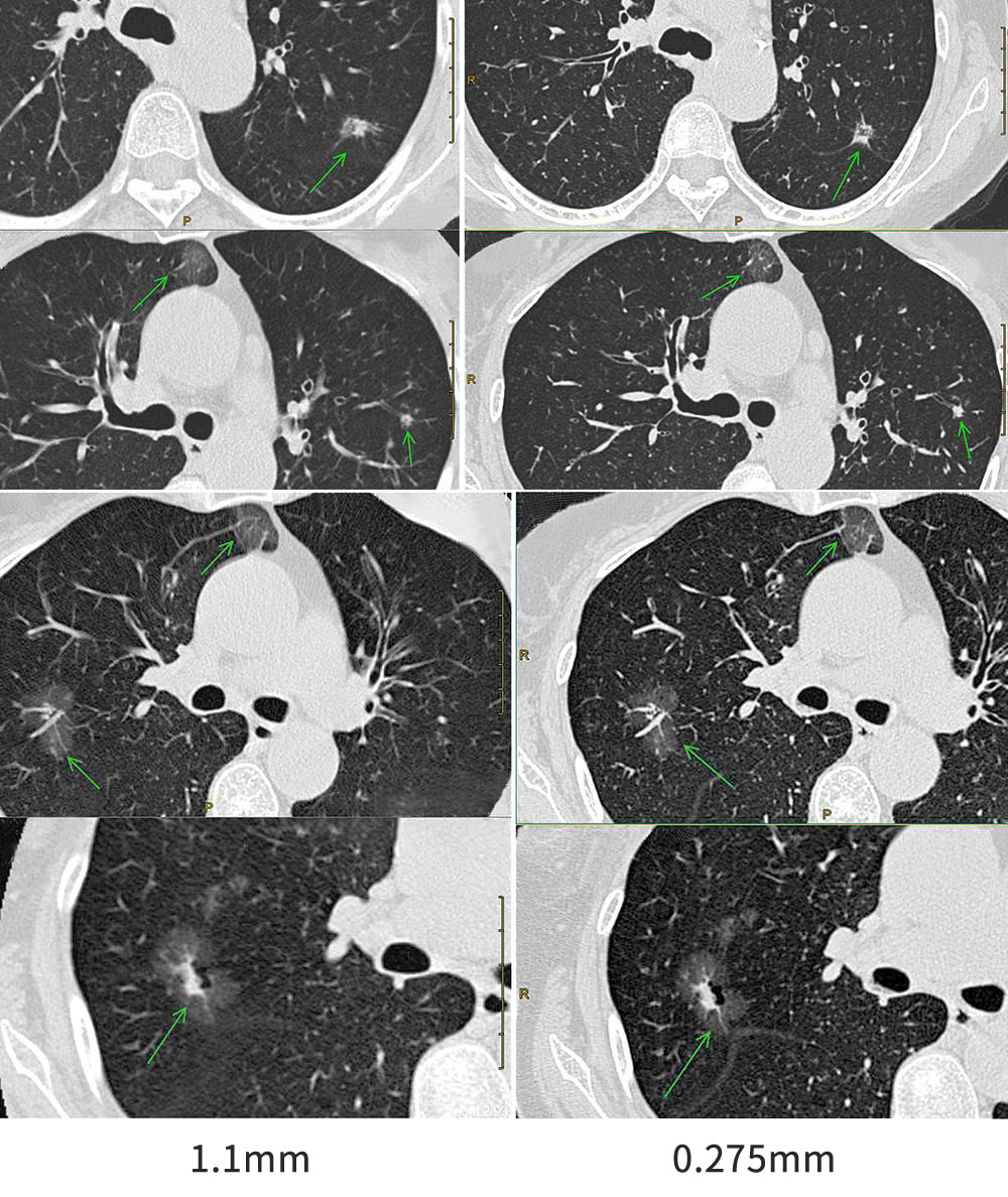
Female, 68 years old, Simultaneous multiple primary lung cancer of both lungs (SMPLC)
Multiple irregular semi-solid patch shadows were seen in both lungs, with solid center and ground glass density shadows around. Burrs and lobulations were seen at the edges, and small bubble transparent areas were seen inside, which were closely related to the adjacent bronchus and the adjacent interlobar pleura was seen to be strained. Thin scanning of 0.275mm showed more lesion definite signs.

Case 3
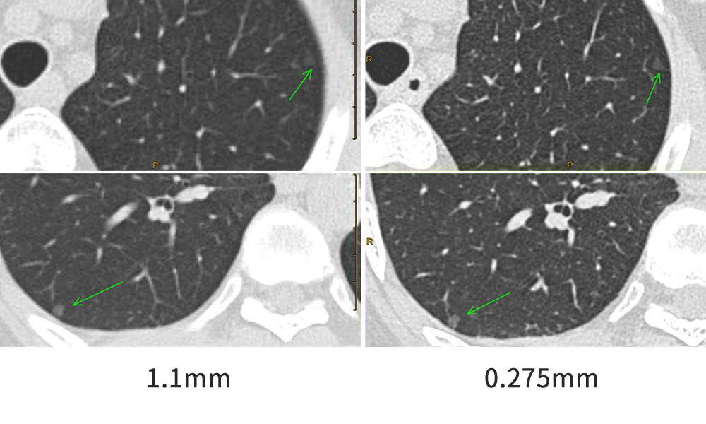
Female, 52 years old, Both lungs have nodules of density of micro ground glass
The relationship between nodules and blood vessels can be clearly shown by 0.275mm precision image. The small nodules with ground glass density and size of 4.5*4.3cm can be seen in the outer base segment of the lower lobe of the right lung (thin Im230). Small ground glass nodules (thin layer 62) are seen in the upper lobe of left lung, about 3.8*3.2mm in size.

Case4
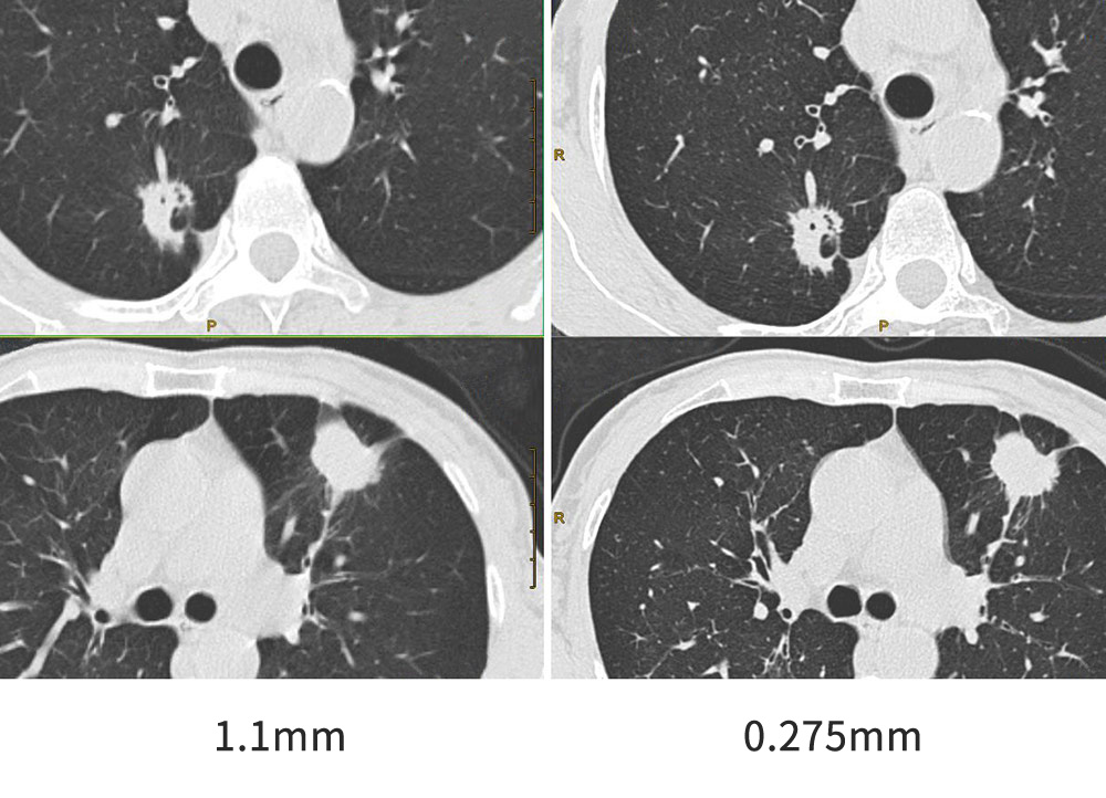
Female, 62 years old, Multiple lesions in both lungs, peripheral lung cancer
Elliptic soft tissue density mass was observed in the anterior segment of the upper lobe of left lung, 2.02cm×2.47cm×2.04cm (left and right, front and back, up and down), average CT value: 52.19Hu, superficial lobulated, short burr, long cable structure connected with the pleura and hilum of the lung. Irregular soft tissue density lesions were observed in the posterior segment of the upper lobe of right lung, and the CT value of the solid part was 44.54Hu, in which multiple vesicles were seen, with lobulation sign, short burrs and long burrs.
