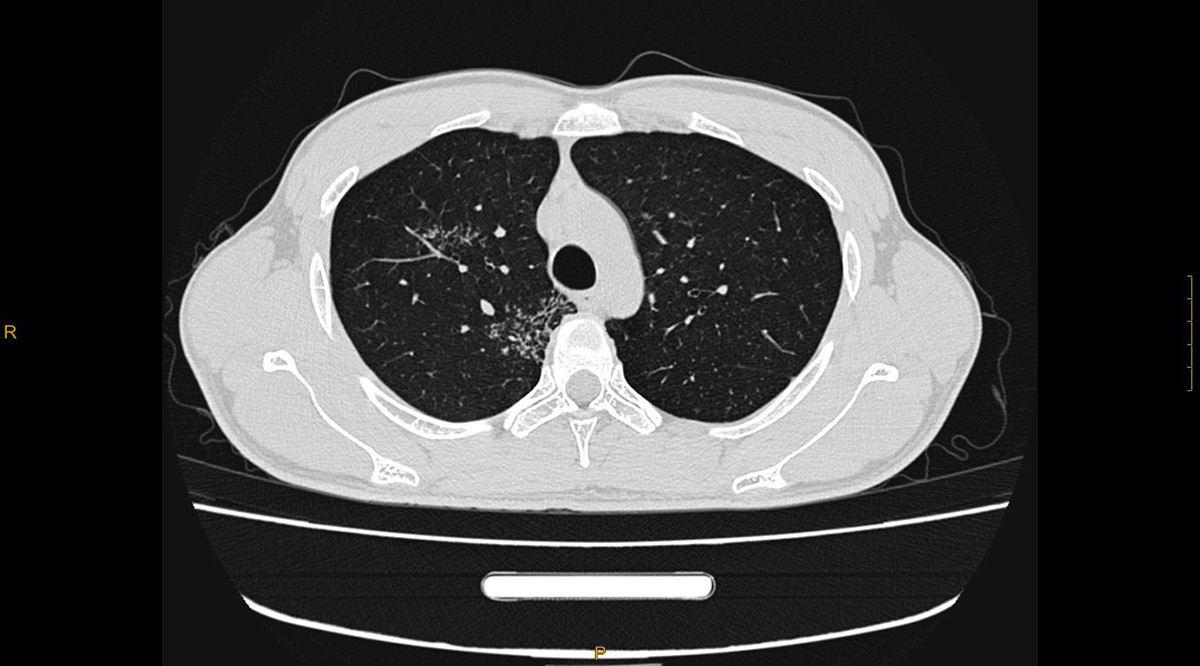
Prof. Ye Maokui Shenyang Campo Imaging Diagnostic Center
Objective: Improve CT diagnostic rate of pulmonary nodules. Method: A total of 100 patients with pulmonary nodules screened by thin slice CT from October 2018 to December 2019 at the author's medical imaging center were followed up as the study subjects, and the benign and malignant imaging characteristics of pulmonary nodules on thin slice CT were analyzed by comparing the pathological results. Result: Of the 100 surgical cases collected, 82 were surgically confirmed as adenocarcinoma of various types, 5 as inflammatory lesions, 2 as sclerosing alveolar cell tumor, 1 as hamartoma, and 10 as benign nodules. Conclusion: The detection rate of lung nodules by thin CT is significantly improved. Radiologists play a decisive role in the differentiation of benign and malignant lung nodules, and fully master the image characteristics of early lung cancer manifested by GGN, so as to achieve early detection, early diagnosis and early treatment of small lung cancer.
As the most common malignant tumor at present, lung cancer has high morbidity and mortality, and the onset age is gradually getting younger. Early detection and early treatment are the most effective methods to reduce the mortality of lung cancer. Many patients are confused and stressed by small pulmonary nodules. Most benign and malignant pulmonary nodules have no specific clinical manifestations and no significant difference in imaging characteristics, which brings difficulties in diagnosis and differential diagnosis. Especially for some small ground glass nodules is one of the difficulties in the differential diagnosis of pulmonary imaging. High-resolution thin layer CT detection of lung nodules can be greatly improved, especially the multiple planar reconstruction (MPR) technology, such as nodules can be viewed from different angles including internal microvessel, fine structure such as bronchiole and lesion edge. The more detailed the information displayed, the smaller the differential range between benign and malignant nodules, and the higher the accuracy of diagnosis. It is very important for early detection, diagnosis, treatment and prognosis of small lung cancer.
The imaging features of 82 cases of small lung cancer confirmed by surgery in author medical image center from October 2018 to December 2019 were summarized. Generally, there is no corresponding clinical symptoms, which is consistent with the clinical characteristics of pulmonary nodules reported in the past. Basically, all cases are discovered accidentally or detected by other hospitals and then consulted by our medical image center after multiple follow-up visits. 22 were male and 66 were female. The youngest age was 34 years, and the oldest was 68 years (Patients older than 70 years were not collected.). All patients underwent low-dose scanning using Precision 32 spectrum CT, independently developed by Liaoning Campo Imaging Medical Co., LTD. Patients were placed in supine position with scanning voltage of 60-70kV, 30-40mA, layer thickness of 5mm, lung window width of 1500, window level of -500, mediastinum window width of 400, window level of 40, scanning range from apex to bottom of lung. Scanning images were sent to the workstation to observe and record the size, shape, number, density and edge of nodules. For some small ground glass nodules, Precision 32 CT was used to scan with ultra-thin layer of 0.275mm, and post-processing techniques such as multi-plane recombination (MPR) were carried out at the same time. Adjust the appropriate window width and window level, focus on observing the morphology and internal fine structure of small nodules from different angles. The image is analyzed and diagnosed by more than two imaging doctors.
Inclusion criteria: Between 35 and 70 years old, there were one or more round or quasi-round lesions in the lung, the diameter of the lesions was no more than 30mm, thin CT scan and reconstruction, after surgical treatment, pathological results were found.
Exclusion criteria: Atelectasis or obvious lymphadenectasis, lung metastases, metastases to the lungs, women in pregnancy and lactation.
A large number of studies and clinical experience concluded that the differentiation of benign and malignant pulmonary nodules mainly includes the size, shape, density, internal and marginal structure and other imaging manifestations of nodules.
Features of benign nodules: It is generally believed that the smaller the pulmonary nodules are, the more regular and benign the nodules are. The larger the nodule and the more irregular the shape, the greater the possibility of malignancy. Small nodules surrounding the lung field, with clear and smooth edge, round, flat, tubular or patchy, and internal calcification or fat density are usually benign. Small nodules with clear boundaries, quasi-circular shape, triangular shape and double convexity are located near the fissures of the lung or within 1.5 cm below the pleura. They can also be connected with interlobular septa or pleura and interlobular fissures. They are usually small lymph nodes in the lung and generally do not need follow-up. Such small nodules may sometimes grow during follow-up, but also tend to be of lymph node origin. Nodules that are located near the lung field and close to the pleura and appear rectilinear on the pleura surface with concave edges suggest contraction or fibrosis. Calcification in nodules is diffuse, stratified, and central, and is often associated with infection (e.g., inflammatory pseudotumor). Popcorn calcification (hamartoma). Fat density is often indicative of hamartoma. Aggregation of micronodules: two or more lesions <10mm, separated from each other, are nodular or glomeratus, suggesting a greater possibility of infective lesions.
Features of malignant nodules: Irregular contour, lobulation, and spicule sign are still common signs of malignant nodules. Deep lobulation has important clinical significance in the diagnosis of malignant nodules. Lobulation refers to the uneven surface edge of tumor, mainly due to the uneven growth rate of tumor in all directions, and also related to the restriction of lung scaffold structure. Especially short spicule signs, are more common in malignant nodules. The proliferation of surrounding pulmonary fibrous connective tissue is mainly caused by tumor cells spreading in all directions or tumor stimulation. The density of pure ground glass nodules increased, suggesting an increased possibility of malignancy, such as microinvasive carcinoma, precancerous lesions, atypical adenomatous hyperplasia. Reactive tumor cells grow along the alveolar wall and replace the alveolar epithelium, the alveolar cavity is not completely filled. Mixed ground glass nodules, the more solid components, the higher the invasiveness, suggesting a greater possibility of malignancy, alveolar collapse and fibrosis caused. The vascularconvergencesign and vascular sign were that the vessels around the nodule gathered to the nodule or went inside the nodule. The vessels were thickened and increased or intertwined into a network. It is the fibrosis and proliferation destruction within the tumor body that lead to the collapse of pulmonary supporting structure pulling on the surrounding blood vessels, or the tumor wrapping and erosion across the blood vessels. Vocule sign is a small unoccluded bronchi or alveoli, usually with cancer cells growing in the form of a wall. Some of the alveolar spaces or bronchi are not filled with tumor cells, and may be stretched by fibrous or scar tissue within the tumor. Pleural indentation refers to the visceral pleura is pulled by the lesion and sags toward the direction of the lesion with arcs on both sides. The degree of malignancy of small nodules is higher when these signs exist simultaneously.